Vertebral and carotid artery dissection
Background
- Primarily categorized as spontaneous vs traumatic/mechanical
- Most frequent cause of CVA in young and middle-aged patients (median age ~40 years)
- Incidence: ~2.6 per 100,000/year for carotid; ~1-1.5 per 100,000/year for vertebral[1]
- Pathophysiology: tear in arterial wall → false lumen formation → luminal stenosis + exposure of subendothelial collagen → local thrombus formation → thromboembolism to intracranial vessels
- Less commonly, dissection of thinner intracranial arteries may lead to SAH through vessel rupture

Key EM Pearls
- Headache/neck pain may precede stroke by up to 14 days — this is the diagnostic window
- Consider in any trauma patient with neurologic deficits despite normal head CT
- Consider in any patient with CVA + neck pain, especially age <50
- Consider in any young patient with Horner syndrome + head/neck pain
- Stroke risk is highest in the first 24 hours after dissection; early diagnosis and treatment are critical
- In the trauma setting, falls under Blunt cerebrovascular injury (BCVI) screening — see Denver screening criteria
Anatomy
- Carotid: internal carotid artery (ICA) is most commonly affected ~2 cm distal to the bifurcation; divided into cervical, petrous, cavernous, and cerebral segments
- Vertebral: arises from the subclavian, runs through the transverse foramina of C6-C2; most vulnerable at C1-C2 (V3 segment) where it is mobile during neck rotation; joins contralateral vertebral to form the basilar artery
Risk Factors
- Any mechanical event involving the neck; symptoms typically occur 2-3 days after event
- Neck trauma (often minor), MVC, falls
- Cervical manipulation (chiropractic, physiotherapy)
- Coughing/sneezing, vigorous exercise, weightlifting, intercourse, roller coasters, yoga
- Connective tissue disease: Ehlers-Danlos syndrome type IV, Marfan syndrome, osteogenesis imperfecta
- History of migraine[2]
- Recent infection (proposed inflammatory vessel wall weakening)
- Hypertension
Clinical Features
Internal Carotid Dissection
- Unilateral headache, face pain, anterior neck pain
- Partial Horner syndrome (miosis and ptosis without anhidrosis) in ~50% — sympathetic fibers run along the ICA
- Cranial nerve palsies — especially CN XII (hypoglossal); also CN IX, X, XI (lower cranial nerves compressed by expanding vessel wall)
- Vision loss (amaurosis fugax, retinal artery occlusion)
- Symptoms of anterior circulation stroke or TIA
Vertebral Artery Dissection
- Posterior neck pain, occipital headache
- May be unilateral or bilateral
- Unilateral facial paresthesia
- Dizziness or Vertigo
- Nausea/vomiting
- Diplopia, nystagmus, and other visual disturbances
- Ataxia
- Dysarthria, dysphagia
- Lateral medullary syndrome (Wallenberg) seen in up to 20% of VAD[3][4]
- Ipsilateral facial pain/numbness, Horner syndrome, ataxia, contralateral body pain/temperature loss, hoarseness, dysphagia, hiccups
- Hemiparesis or quadriparesis — may progress rapidly to locked-in syndrome (basilar involvement)
Differential Diagnosis
Neck Trauma
- Penetrating neck trauma
- Blunt neck trauma
- Cervical injury
- Neurogenic shock
- Spinal cord injury
Other considerations:
- Migraine — most common misdiagnosis; but new-type headache in a patient without migraine history warrants imaging
- Cervical strain / musculoskeletal neck pain — absence of neurologic symptoms does NOT exclude early dissection
- Subarachnoid hemorrhage — thunderclap headache; intracranial dissection can cause SAH
- Cerebellar stroke from other embolic sources
- Vertebrobasilar insufficiency
- Meningitis — neck pain + headache; check for fever, meningismus
- Giant cell arteritis (patients >50 with new headache)
- Cavernous sinus thrombosis — if cranial nerve palsies with proptosis
Evaluation
Workup
- CT head (non-contrast): obtain first to assess for concomitant SAH, intracranial hemorrhage, or established infarct
- A normal head CT does NOT exclude dissection
- CTA head and neck (aortic arch through circle of Willis): imaging modality of choice
- Sensitivity/specificity ~95-98% with modern ≥16-slice CT[5]
- CTA has been shown to be equivalent to MRA
- Look for: luminal narrowing ("string sign"), intimal flap, intramural hematoma (crescent sign), pseudoaneurysm, occlusion, "flame-shaped" tapering
- MRI/MRA with fat-suppressed T1: shows intramural hematoma as hyperintense crescent; preferred if already obtaining MRI for stroke workup; better for follow-up imaging
- MRI brain (DWI): most sensitive for acute posterior fossa ischemia (CT misses many small cerebellar/brainstem strokes)
- Labs: CBC, BMP, coags, troponin, glucose, type and screen
- ECG: baseline; stroke can cause ECG changes (deep TWI, prolonged QT)
- Doppler ultrasound: insufficient — misses skull base segments; a negative ultrasound does NOT exclude dissection
Screening in Trauma
- Apply the Denver screening criteria for BCVI screening in blunt trauma patients
- If positive findings on screening → obtain CTA
Denver screening criteria for blunt cerebrovascular injury
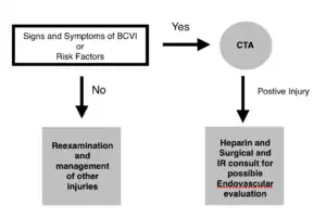
The Denver Screening Criteria are divided into risk factors and signs and symptoms
Signs and Symptoms
- Arterial hemorrhage
- Cervical bruit
- Expanding neck hematoma
- Focal neurologic deficit
- Neuro exam inconsistent with head CT
- Stroke on head CT
Diagnosis
- Confirmed by CTA or MRA showing vessel wall abnormality (intimal flap, intramural hematoma, stenosis, pseudoaneurysm, occlusion)
- Biffl Grading Scale (for traumatic BCVI; determines management):
| Grade | Injury | Management |
|---|---|---|
| I | Intimal irregularity or dissection with <25% luminal narrowing | Antithrombotic therapy (ATT) |
| II | Dissection/intramural hematoma with ≥25% luminal narrowing, intraluminal thrombus, or raised intimal flap | ATT; follow-up imaging at 7-10 days; endovascular if progresses |
| III | Pseudoaneurysm | ATT ± endovascular repair |
| IV | Complete occlusion | ATT ± endovascular repair |
| V | Transection with active extravasation | Emergent hemorrhage control (surgery/endovascular) |
- Many grade I-II injuries heal spontaneously within 7-10 days with antithrombotic therapy
- Intracranial extension changes management — higher SAH risk; anticoagulation may be contraindicated
Management
Acute Stroke Management
- Activate stroke team — do not delay
- IV alteplase (tPA): dissection-related stroke is NOT a contraindication to thrombolysis within standard time windows per AHA/ESO guidelines[6]
- Do NOT give if dissection is intracranial — risk of SAH or ICH
- Do NOT give if aortic dissection involved — may worsen dissection
- Otherwise, give per same guidelines as ischemic CVA
- In meta-analysis, thrombolytics appear equally efficacious in dissection-related stroke vs stroke from other causes[7]
- Mechanical thrombectomy: consider for large vessel occlusion (basilar artery, proximal MCA) per standard stroke protocols
- Obtain emergent vascular surgery or neurointerventional consult for possible endovascular intervention
Antiplatelet vs Anticoagulation Therapy
- CADISS trial (2015) and TREAT-CAD trial (2021): no significant difference between antiplatelets vs anticoagulation in preventing stroke/death after cervical artery dissection[8][9]
- Choice is guided by location and clinical scenario:
- Heparin → warfarin (anticoagulation): if dissection is extracranial AND causes neurologic deficit[10]
- Aspirin: if dissection is intracranial — anticoagulants avoided due to weaker medial walls and higher SAH risk[11]
- Aspirin: if dissection is extracranial with no neurologic deficit (stroke prevention)
- Aspirin: if large completed infarct (risk of hemorrhagic transformation with anticoagulation)
- Aspirin: preferred in polytrauma or other active bleeding concerns
- Practical summary: when in doubt, aspirin is the safer choice — the evidence shows no superiority of anticoagulation, and aspirin carries less bleeding risk
- If tPA was given, wait 24 hours before starting antithrombotic therapy
- Do not anticoagulate if NIHSS ≥15 (high risk of hemorrhagic transformation)
- Duration: typically 3-6 months; follow-up imaging (CTA or MRA) guides discontinuation; ~90% of dissections heal in this period
Endovascular/Surgical Intervention
- Emergent for Grade V (transection with hemorrhage)
- Consider for Grade II-IV injuries that progress despite antithrombotic therapy
- Consider if antithrombotic therapy is contraindicated (e.g., concurrent intracranial hemorrhage, solid organ injury)
- Stenting is increasingly used but data on superiority over ATT alone remains limited
Complications
- CVA: risk of stroke is highest in the first 24 hours after dissection; remains elevated for first 2 weeks
- SAH: if dissection extends intracranially (especially vertebral V4 segment)
- Recurrent dissection: ~1% annual risk; higher in those with connective tissue disorders or family history
- Pseudoaneurysm formation: may require long-term follow-up and possible intervention
Disposition
- Admit all confirmed or strongly suspected cervical artery dissections
- Symptomatic patients (headache/neck pain only, no stroke): still admit — stroke risk is highest in first 2 weeks; serial neurologic examinations
- Start antithrombotic therapy in the ED — do not defer to the floor
- Follow-up imaging: CTA or MRA at 7-10 days (traumatic/BCVI) or 3-6 months (spontaneous) to assess healing
- Counsel:
- Avoid cervical manipulation (chiropractic, aggressive physiotherapy) — indefinitely
- Seek immediate care if new neurologic symptoms develop
- Medication compliance with antithrombotic therapy
- Annual recurrence risk ~1% after initial episode; higher with connective tissue disorders
- Return precautions: any new weakness, numbness, speech difficulty, dizziness, imbalance, visual changes, severe worsening headache
See Also
- Blunt cerebrovascular injury
- Denver screening criteria
- Blunt neck trauma
- Penetrating neck trauma
- Ischemic stroke
- Stroke (main)
- CVA (tPA)
- Thrombolytics for acute ischemic stroke
- Cerebellar stroke
- Vertebrobasilar insufficiency
- Subarachnoid hemorrhage
- Horner syndrome
- Lateral medullary syndrome
- Alteplase
- Anticoagulation
- Antiplatelet
- Heparin
- Aspirin
- TIA
- Vertigo
- Neck pain
- Cervical fractures and dislocations
- Strangulation
External Links
- StatPearls — Vertebral Artery Dissection
- StatPearls — Vertebral Artery Injury
- Medscape — Vertebral Artery Dissection Treatment & Management
- AJNR — Blunt Cerebrovascular Injuries: Advances in Screening, Imaging, and Management (2021)
- Scand J Trauma — Best practice guidelines for BCVI (2018)
- EAST — BCVI Practice Management Guideline (2020)
References
- ↑ Vertebral Artery Dissection. StatPearls. NCBI. 2025.
- ↑ De Giuli V et al. Association Between Migraine and Cervical Artery Dissection: The Italian Project on Stroke in Young Adults. JAMA Neurol. Published online March 6, 2017. doi:10.1001/jamaneurol.2016.5704
- ↑ Lee MJ, Park YG, Kim SJ, Lee JJ, Bang OY, Kim JS. Characteristics of stroke mechanisms in patients with medullary infarction. Eur J Neurol. 2012;19(11):1433-1439.
- ↑ Kim JS. Pure lateral medullary infarction: clinical-radiological correlation of 130 acute, consecutive patients. Brain. 2003;126(Pt 8):1864-1872.
- ↑ Provenzale JM, Sarikaya B. Comparison of test performance characteristics of MRI, MR angiography, and CT angiography in the diagnosis of carotid and vertebral artery dissection: a review of the medical literature. AJR Am J Roentgenol. 2009 Oct;193(4):1167-74. doi: 10.2214/AJR.08.1688. PMID: 19770343.
- ↑ Zinkstok SM, Vergouwen MD, Engelter ST, et al. Safety and functional outcome of thrombolysis in dissection-related ischemic stroke: a meta-analysis of individual patient data. Stroke. 2011;42:2515–2520.
- ↑ Zinkstok SM, Vergouwen MD, Engelter ST, Lyrer PA, Bonati LH, Arnold M, Mattle HP, Fischer U, Sarikaya H, Baumgartner RW, Georgiadis D, Odier C, Michel P, Putaala J, Griebe M, Wahlgren N, Ahmed N, van Geloven N, de Haan RJ, Nederkoorn PJ. Safety and functional outcome of thrombolysis in dissection-related ischemic stroke: a meta-analysis of individual patient data. Stroke. 2011 Sep;42(9):2515-20. doi: 10.1161/STROKEAHA.111.617282. Epub 2011 Jul 28. PMID: 21799165.
- ↑ CADISS trial investigators; Markus HS, Hayter E, Levi C, Feldman A, Venables G, Norris J. Antiplatelet treatment compared with anticoagulation treatment for cervical artery dissection (CADISS): a randomised trial. Lancet Neurol. 2015 Apr;14(4):361-7. doi: 10.1016/S1474-4422(15)70018-9. Epub 2015 Feb 12. Erratum in: Lancet Neurol. 2015 Jun;14(6):566. PMID: 25684164.
- ↑ Engelter ST, Traenka C, Gensicke H, et al. Aspirin versus anticoagulation in cervical artery dissection (TREAT-CAD): an open-label, randomised, non-inferiority trial. Lancet Neurol. 2021;20(5):341-350.
- ↑ Vasc Med. 2011 Feb;16(1):35-77. doi: 10.1177/1358863X11399328. Erratum in: Vasc Med. 2011 Aug;16(4):317. PMID: 21471149.
- ↑ Debette S, Mazighi M, Bijlenga P, et al. ESO guideline for the management of extracranial and intracranial artery dissection. Eur Stroke J. 2021 Sep;6(3):XXXIX-LXXXVIII. doi: 10.1177/23969873211046475. PMID: 34746432; PMCID: PMC8564160.